Deoxyribonucleic acid (DNA) is the basic hereditary material found in the nucleus of most cells. This genetic information is passed on from one generation to the next and is required for protein synthesis. This important life information is packaged in the nucleus in a highly structured and organised manner. DNA is tightly wrapped around proteins called histones and this complex structure is the nucleosome. Nucleosomes are the structural and functional units of a chromatin. These are then folded and looped over other proteins and this chromatin is further compressed into the chromatid of a chromosome.
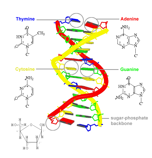
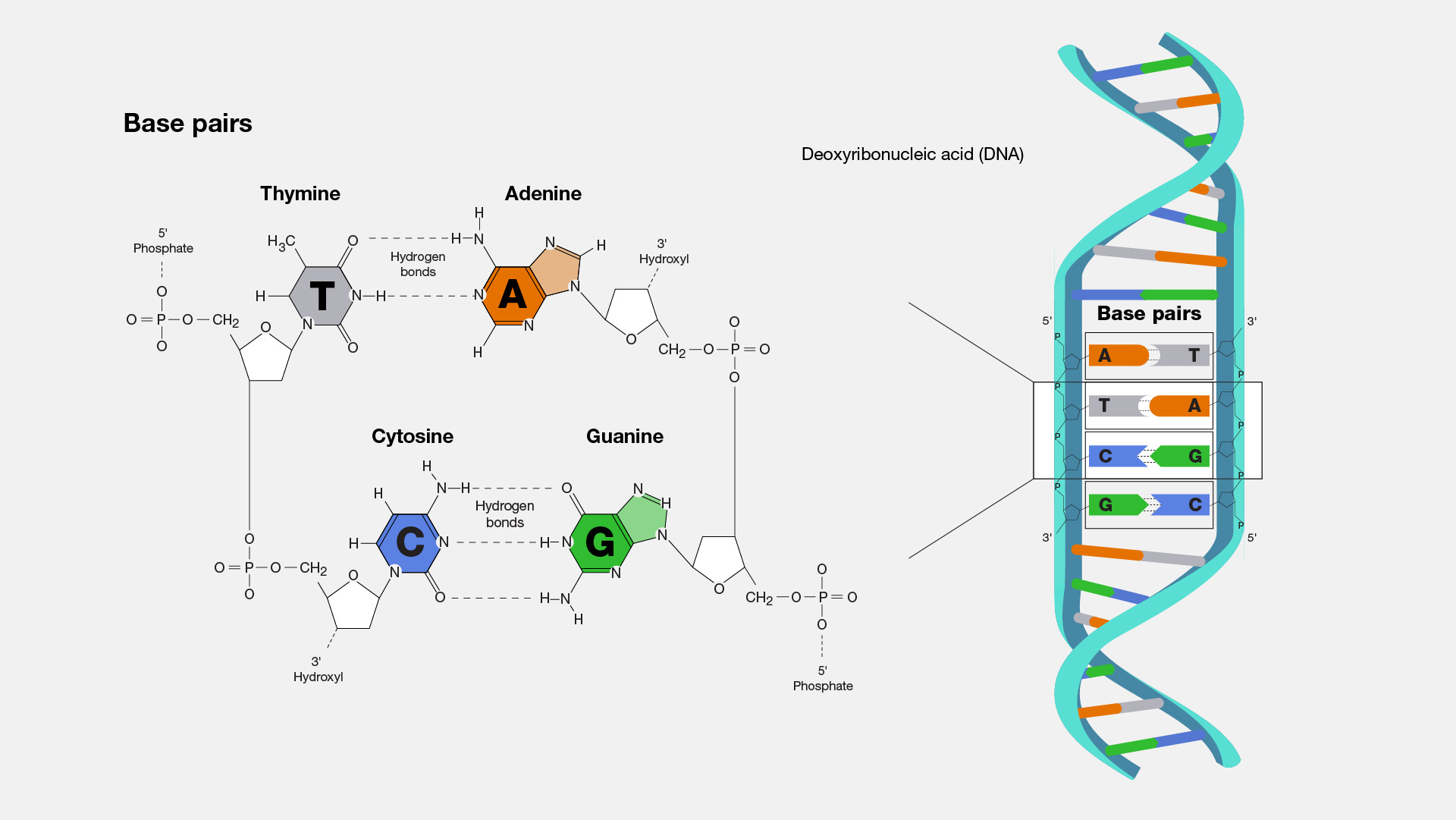
DNA is a linear polymer that is made up of nucleotide units. The nucleotide unit consists of a base, a deoxyribose sugar, and a phosphate. There are four types of bases: adenine (A), thymine (T), guanine (G), and cytosine (C). Each base is connected to a sugar via a ß glycosyl linkage. The nucleotide units are connected via the O3' and O5' atoms forming phosphodiester linkages.In normal DNA, the bases form pairs: A to T and G to C. This is called complementarity. A duplex of DNA is formed by two complementary chains that are arranged in an anti-parallel manner.
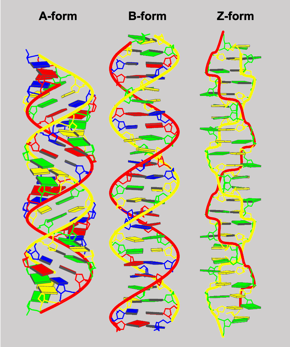
DNA can have several conformations. The most common one is called B-DNA. B-DNA is a right-handed double helix with a wide and narrow groove. The bases are perpendicular to the helix axis. DNA can also be found in the A form in which the major groove is very deep and the minor groove is quite shallow. A-DNA is also a right-handed double helix. A very unusual form of DNA is the left-handed Z-DNA. In this DNA, the basic building block consists of two nucleotides, each with different conformations. Z-DNA forms excellent crystals.
Ribonucleic acid (RNA) is present in several forms - messenger RNA (mRNA), transfer RNA (tRNA), and ribosomal RNA (rRNA). Each of these RNA forms is involved in different steps of protein synthesis. mRNA is generated from DNA and is the template for protein synthesis. tRNA is critical to the translation of the mRNA sequence into protein sequence. rRNA are components of the ribosomes which are the sites of protein synthesis.
The mRNA once produced, comes out of the nucleus and meets with the ribosome. The smaller and bigger subunits of the robosome locks on to the mRNA and reads the information from the mRNA. The ribosomes pick the appropriate t-RNA which has brought the corresponding aminoacid to the three letter code found on the mRNA to make the protein. This last step is repeated until the stop codon is reached and the protein synthesis is completed.
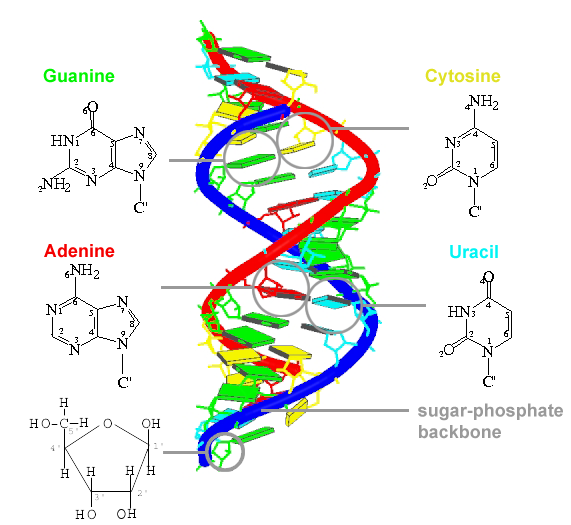
RNA is a polymer that contains ribose sugar instead of the deoxyribose sugars found in DNA. The normal base composition is adenine, uracil, guanine, and cytosine.
RNA can form double stranded duplexes. These duplexes are in the A conformation because the 2'OH precludes the B conformation. More commonly, RNA is single stranded and can form complexes and unusual shapes. One example is tRNA which contains about 70 bases that are folded such that there are base paired stems and open loops. The completely folded tRNA is L shaped. Another very interesting type of RNA is called a ribozyme, which is an RNA that has a catalytic activity.
The right-handed double-helical Watson-Crick Model for B-form DNA is the most commonly known DNA structure. In addition to this classic structure, other forms of DNA have been observed. In A-form DNA, the ribose sugar adopts 3'-endo conformation, whereas in B-form DNA, the sugar conformation is 2'-endo. A second major difference between A- and B-forms is the placement of the base-pairs within the duplex. In B-form, the base-pair plane is almost centered over and perpendicular to the helical axis, but in A-form the plane is both tilted and displaced away from the helical axis towards the major groove. Z-form DNA is a left-handed zig-zag that can form when an alternating purine-pyrimidine sequence is present such as GCGCGC. The helical structure of DNA is thus variable and depends on the sequence as well as the environment.
Shown: DNA duplexes in A-form, B-form, and Z-form, generated using ideal fiber parameters. Download links are provided for model coordinates (PDB format). Source: Web-x3DNA fiber building tool.
| form | rise/unit(Å) | twist/unit(°) | #bp/unit | download |
|---|---|---|---|---|
| A | 2.548 | 32.7 | 1 | A-form model |
| B | 3.375 | 36.0 | 1 | B-form model |
| Z | 7.250 | -60.0 | 2 | Z-form model |